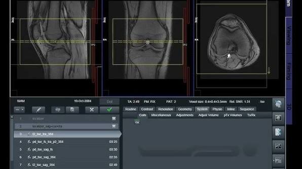
MRI Musculoskeletal (MSK) Imaging

Categories: MRI Technology
Wishlist Share

About Course

Learn the fundamentals of musculoskeletal (MSK) MRI, including anatomy, patient positioning, routine protocols, imaging planes, sequence selection, and image planning for common MSK examinations. This course provides practical guidance for producing high-quality MRI images of the shoulder, knee, wrist, hand, ankle, and foot.


Course Content

MSK Anatomy & Positioning
Learn musculoskeletal anatomy, MRI positioning, anatomical landmarks, imaging planes, and patient setup for common MSK examinations.

  • MSK Anatomy & Positioning
  • Shoulder Anatomy & MRI Positioning — Quiz

MSK Anatomy: Major Joints & Soft Tissues
Introduction Musculoskeletal (MSK) MRI evaluates bones, joints, muscles, tendons, ligaments, cartilage, and other soft tissues. Understanding normal anatomy helps the MRI technologist select appropriate positioning, imaging planes, and coverage. Major Joints Shoulder Humeral head Glenoid Rotator cuff tendons Labrum Acromioclavicular (AC) joint Long head of the biceps tendon Elbow Distal humerus Proximal radius and ulna Common flexor and extensor tendons Ulnar collateral ligament Radial collateral ligament Wrist Distal radius and ulna Carpal bones Triangular fibrocartilage complex (TFCC) Flexor and extensor tendons Scapholunate ligament Hip Femoral head and neck Acetabulum Labrum Articular cartilage Gluteal tendons Iliopsoas tendon Knee Femur Tibia Patella Menisci Anterior cruciate ligament (ACL) Posterior cruciate ligament (PCL) Medial and lateral collateral ligaments Articular cartilage Ankle Distal tibia and fibula Talus Achilles tendon Medial and lateral ankle ligaments Articular cartilage Major Soft Tissues Muscles: MRI can evaluate muscle size, signal, tears, edema, and other abnormalities. Tendons: Tendons connect muscles to bones and are commonly evaluated for tendinopathy and tears. Ligaments: Ligaments stabilize joints and can be assessed for sprains and tears. Cartilage: Articular cartilage covers the joint surfaces and can be evaluated for defects, degeneration, and loss. Labrum: Fibrocartilage structures such as the shoulder and hip labrum help stabilize the joint. MRI Positioning Principles Proper positioning is important for obtaining diagnostic images. The technologist should: Center the anatomy appropriately within the MRI coil. Use the appropriate dedicated coil whenever available. Immobilize the patient to reduce motion. Select imaging planes based on the anatomy and clinical indication. Ensure adequate anatomical coverage. Maintain MRI safety throughout the examination. Quick Review Shoulder → Elbow → Wrist → Hip → Knee → Ankle Remember the major structures evaluated in MSK MRI: Bones → Muscles → Tendons → Ligaments → Cartilage → Labrum → Soft tissues Learning Objective After completing this lesson, the learner should be able to identify major MSK joints and recognize the primary bones, tendons, ligaments, cartilage, and soft-tissue structures commonly evaluated with MRI.

Student Ratings & Reviews

No Review Yet
No Review Yet